The Future of Medical Imaging
The World Health Organization states medical imaging equipment is the most crucial tool doctors, nurses and other medical staff use to help patients.
This wide variety of equipment, including MRI scanners, CT scanners, PET scanners, and X-ray machines, assists in preventative care as well as palliative care. The machines can provide correct diagnoses, assess disease status, and provide general information for a well-trained, interdisciplinary team of professionals.
Substantial advances in medical imaging technology are now taking place, both in software such as cloud computing tools and hardware such as medical imaging cable assemblies. Looking at future trends can offer a glimpse at the demands the medical device industry may soon see.
MRI
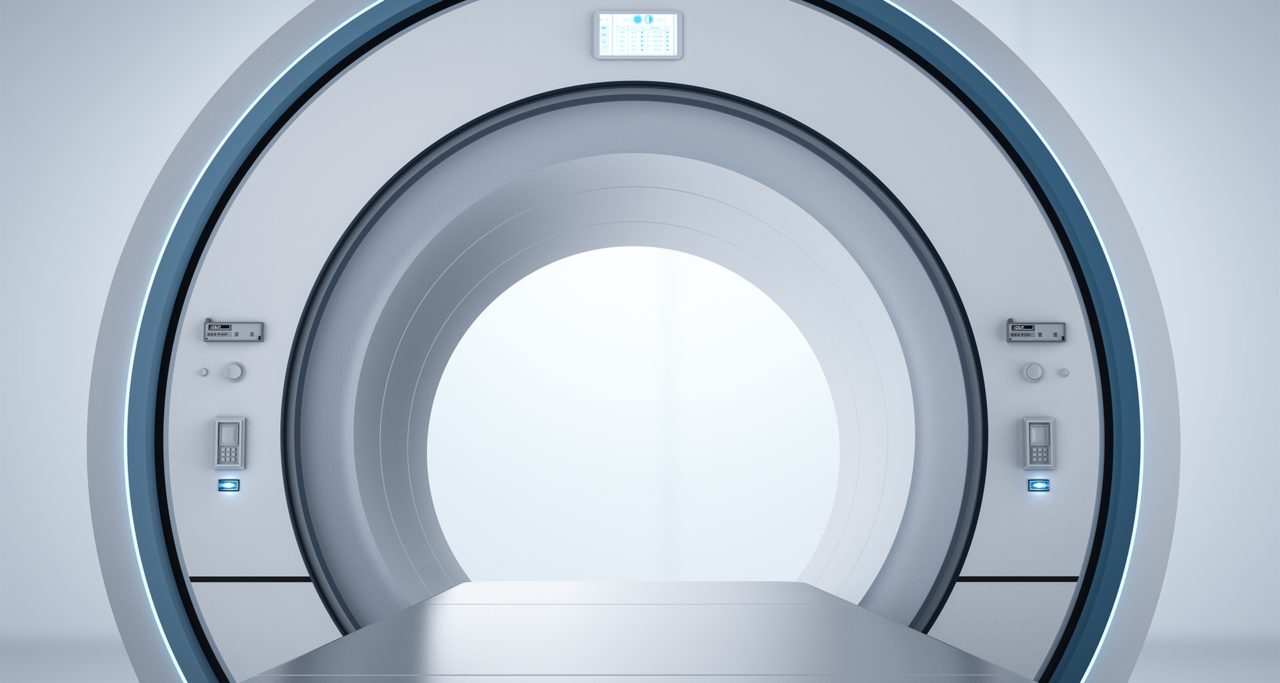
For musculoskeletal disorders, the MRI scanner is the diagnostic tool of choice throughout the world. These machines require many complex and delicate parts, including the MRI cable assembly, along with advanced programs for running the entire system. Current and future advances include new technology that allows for increased usage and improved patient experiences.
New MRI Uses
Previously, doctors and technicians have had trouble administering MRIs to patients with lung or heart conditions. The short relaxation time of the muscular tissue and long exam times made it a less effective imaging option.
Recently, however, technology has improved sequencing, reducing scan times. 3-D chest volume scans are also now possible, allowing for expanded cardiac and pulmonary diagnostic and monitoring opportunities.
The FDA has just cleared the use of MRI scans for noninvasive prostate exams as well. As a result, manufacturers have improved resolution and signal-to-noise ratio in these procedures.
Some companies are also experimenting with scanning technology that will tilt patients up to 90 degrees. This permits the imaging of weight-bearing body parts that otherwise would not be possible using supine or prone methods.
Minimized Scan Times
Another recent improvement in MRI imaging is reduced time to perform scans. Shorter durations mean patients are more comfortable, and people are able to spend less time at medical appointments. Lower scan times also allow technicians to use MRI more frequently as a diagnostic tool in challenging patients, including those who suffer from anxiety .
Stronger and Quieter
The FDA recently allowed the use of a 7-Tesla MRI system, which greatly improves the overall quality of imaging. This new system doubles the strength of an MRI scanner’s static magnetic field. The increased power will result in expanded MRI uses—specifically regarding musculoskeletal and neurological conditions.
In addition to this, acoustic noise, long a major complaint, has been reduced. This will allow noise-sensitive patients to take advantage of MRI technology.
CT Scans
CT scans, or computed tomography scans, are crucial diagnostic and monitoring tools in emergency medical systems. This type of scan involves X-ray tubes, connected by a CT scan cable assembly, which create an image of a patient using digital detectors. Current and future trends in this medical imaging technology include both equipment and software advancements.
Expanded Use of Spectral Imaging
More challenging patients, including those who are obese or have complex tumor or lesion growths, are now able to benefit from CT scans. This is thanks to gemstone spectral imaging, or GSI. This new technology offers reduced patient exposure to radiation as well as improved diagnostics through connections with other available tools.
With improved contrast images, CT scans will not be limited to identifying bone and joint problems. They can also be used for spotting more subtle signs of disease.
Improved Accuracy
New trends are helping CT scans become more accurate as well, improving disease management. Equipment upgrades allow for imaging without the use of sedation thanks to a reduction in time needed for scans. Improved accuracy and shortened scan times aren’t achieved at the expense of adherence to XR-29 standards and the maintenance of safe dosing levels throughout procedures.
Trends in Safety
Thanks to advancements in cardiac imaging, medical professionals are trending toward the use of CT technology for angiography. By using this noninvasive method first, less time is needed for many catheterizations and nuclear perfusion scans.
New improvements in computers have additionally allowed for better image resolution. By having better iterations of an image, patients require a lower, less risky radiation dose.
PET
Positron emission tomography (PET) scans date back to 1972. They remain a common and important imaging technology for monitoring and diagnosing disease. PET machines, which rely on a PET cable assembly and other high-performance connective technology, use radioactive tracers for early detection of diseases including cancer, heart conditions, and neurological concerns. New uses and more affordable imaging are all part of future and current trends in PET.
Neurodegenerative Uses
While most doctors will prescribe a PET scan to diagnose and monitor a cancer occurrence, this technology is moving toward detecting neurodegenerative disease. New radiotracers are available for the medical industry, allowing this tool to be used in Alzheimer’s disease treatment.
More Cost-Effective Systems
PET scans are becoming more affordable, which is important as cost is frequently a barrier for patients who need care. There are a variety of new scanners that are priced competitively and are focused on single-organ usage.
While there are more specialized scanners, clinical technicians now have access to new technology that combines PET and MRI systems. This reduces overhead and provides improvements in the ease and accuracy of medical imaging.
X-Rays
Scientific American described the importance of X-ray technology back in 1916, when sharing a new process for removing bullets. While X-ray machines create the same rays of electromagnetic radiation, today’s improved equipment has modernized the technology. Updates to everything from the X-ray cable assembly to new techniques are the basis for current and future trends.
3-D Imaging
With a new imaging technique called ghost imaging, researchers are studying the potential for using X-ray technology to create 3-D images for mammography. Previously, X-rays were of limited function in this field due to the concerns regarding radiation. But 3-D ghost imaging requires significantly smaller doses and has more options for use.
More Options for Assembly
Today, X-ray technology varies dramatically in terms of resolution, power, magnification, and detector quality. Some machines are now utilizing copper wire instead of the traditional gold wire as a medical imaging cable assembly. This can result in smaller, more affordable devices.
Trends in medical imaging are moving towards reduced times, better quality, and more diagnostic options for the field as a whole. This is great news for both patients and professionals in the industry.
Back To Blog